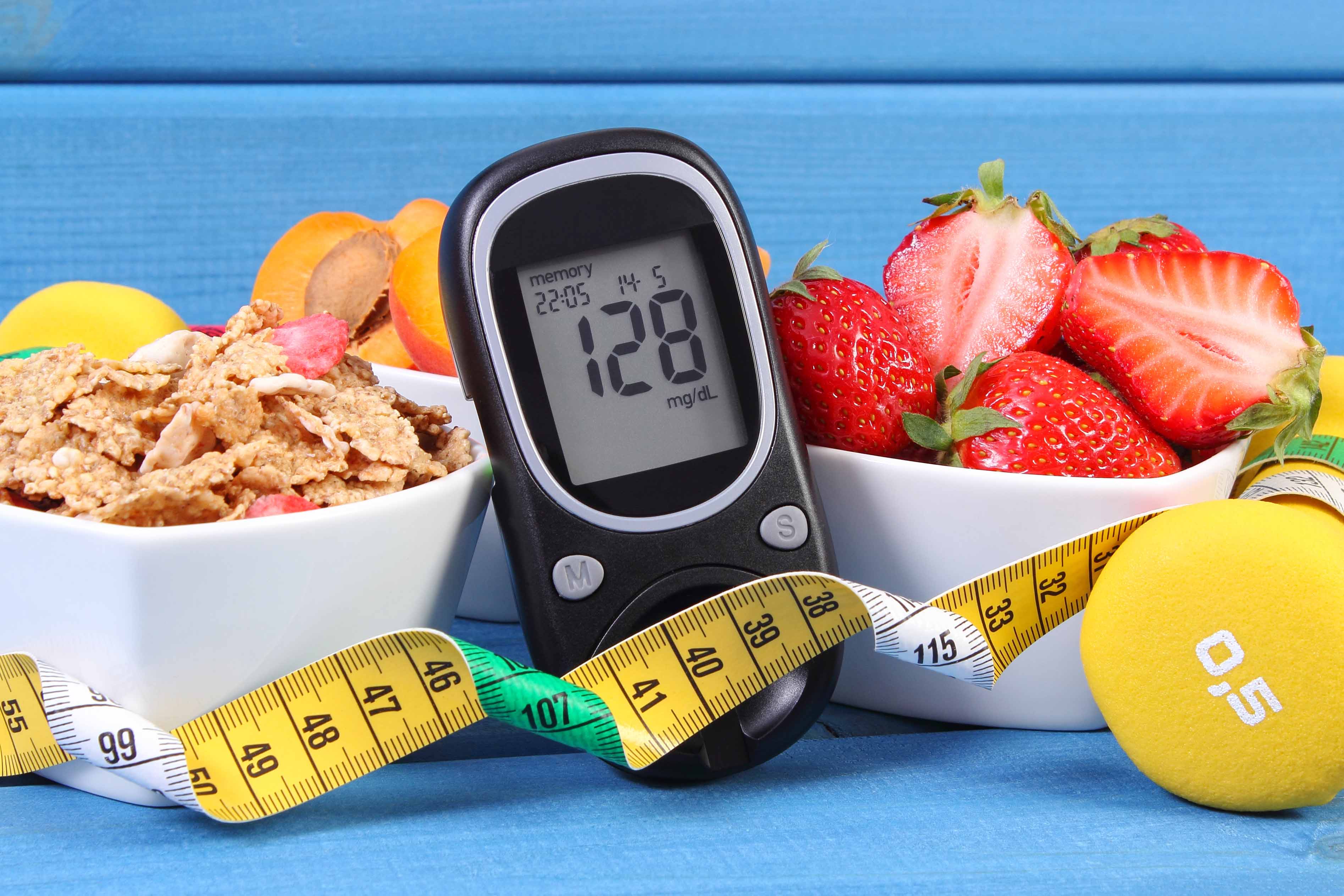
The increase of adiposity after menopause has been associated with an increased risk of breast cancer. In truth, one of the keys may lie in hyperinsulinism.
Diabetes/insulin resistance and breast cancer are pathologies that are quite different but the route of insulin has a central, common role in both. By identifying this metabolic alteration, we will be able to act using our best weapons: diet, exercise, and metformin.
Dr. Celia Gonzalo Gleyzes – Neolife Medical Team
About insulin
Insulin is a peptide hormone produced by the beta cells of the pancreas when there is an increase of plasma glucose (in the blood).
This hormone stimulates glucose uptake by the muscles and adipose tissue. In the liver, insulin inhibits gluconeogenesis (the formation of glucose) and the release of glucose.

High levels of glucose make the liver and the muscles store the excess glucose.
In the adipocytes, insulin promotes the transport of fatty acids in the bloodstream, their storage (lipogenesis), and it inhibits lipolysis. Summing up, insulin facilitates the storage of energy so that it can be mobilized when the levels of insulin are low (fasting).
Apart from the aforementioned organs, insulin also has effects on the neurons, the endothelial cells (cells that line the blood vessels), and immune cells.
To avoid getting lost in the different concepts, we’re going to briefly review the route of insulin.
Insulin attaches to the insulin receptor and activates cellular signaling pathways that are key regulators of cellular homeostasis. These signaling pathways are altered in the majority of biologically aggressive cancers.
Under circumstances rich in nutrients, insulin is released and attaches to the insulin receptor. This binding of insulin promotes tyrosine phosphorylation of the insulin receptor and the insulin receptor substrate (IRS). The IRS, in turn, phosphorylates phosphatidylinositol-3-kinase (PI3K) and activates signaling of the AKT/mTOR pathway.
Insulin also activates signaling of insulin/IGF-1. IGF-1 binds to its receptor, leading to phosphorylation cascades that activate the PI3K/AKT/mTOR pathway and the ras/RAF/mitogen-activated protein kinase (MAPK).
Insulin resistance
Overeating alters the balance between energy storage and energy consumption, leading the individual to prediabetes (insulin resistance) and later to type 2 diabetes mellitus. Having high levels of insulin because of that overeating brings about desensitization of the skeletal muscle. The pancreas is forced to produce insulin continuously and these beta cells will eventually die (the stage in which the person with type 2 diabetes mellitus will require insulin).
Hyperinsulinism in the liver causes dyslipidemia (increased LDL cholesterol and triglycerides) and favors the development of fatty liver (hepatic steatosis).
In the brain, a sustained increase in insulin stimulates appetite, favoring weight gain. In the skeletal muscle, hyperinsulinism and insulin resistance make the muscle unable to take advantage of glucose; thus, the person affected will have less tolerance to physical exercise and also general tiredness.
In adipose tissue, hyperinsulinism increases lipid accumulation and causes inflammation.
In blood vessels and the kidney, insulin promotesendothelial cell damage and renal dysfunction due to increased nitric oxide synthesis, increased reactive oxygen species, and decreased cell adhesion.
Insulin and cancer
Below, we will note the mechanisms that relate hyperinsulinism and tumorigenesis:
- Glycolysis: the PI3K/AKT/mTor signaling pathway regulates glucose metabolism and aerobic glycolytic metabolism. Aggressive cancers are glucose dependent and produce much of their energy through aerobic glycolysis (Warburg effect) and not through the Krebs cycle (mitochondrial oxidative phosphorylation).
- Oxidative stress: reactive oxygen species (ROS) are increased. These can damage DNA, trigger cancer, or favor its progression.
- Cellular mobility: through the activation of the MAPK and PI3K/AKT pathways. Modulation of cytoskeleton proteins occurs, including vimentin and actin.
- Epithelial-mesenchymal transition (EMT).
- Inflammation: due to an increase in inflammatory cytokines, which – among other effects – may favor the formation of new vessels (angiogenesis). In addition, activation of M2 macrophages causes them to secrete epithelial growth factor (EGF) and tumor growth factor beta (TGFbeta), both involved in invasion, metastasis, and cell turnover. Activation of these pathways is related to a poor prognosis in triple-negative breast cancer.
- Angiogenesis: insulin activates pathways that increase the production of vascular growth factors.
Prevention and treatment of insulin resistance: taking metformin
Metformin is a widely prescribed, oral hypoglycemic drug. It is the first line of treatment in type 2 diabetes and its use is also approved in the treatment of polycystic ovary syndrome and gestational diabetes. Metformin is tolerated fairly well, although some people may experience diarrhea, nausea, and epigastric pain.
This drug inhibits hepatic glucose production, increases insulin sensitivity, and decreases intestinal glucose absorption – all favoring the decrease of circulating insulin. Its anticancer potential would be explained precisely by that.
Upon detection of insulin resistance, fasting hyperinsulinism, prediabetes, or type 2 diabetes mellitus, it is essential for us to begin to act through dietary control, calorie restriction, physical activity, and – if necessary – the use of metformin.
The article cited focused on breast cancer, but these mechanisms would also be involved in other types of cancer. Oncologists should monitor this metabolic alteration closely as it could prevent the appearance of cancers, improve patients’ evolution, and prevent recurrences (1).
BIBLIOGRAPHY
(1) https://www.ncbi.nlm.nih.gov/pmc/articles/PMC7045050/ Yee, Lisa D et al. “Metabolic Health, Insulin, and Breast Cancer: Why Oncologists Should Care About Insulin.” Frontiers in endocrinology 11 58. 20 Feb. 2020, doi:10.3389/fendo.2020.00058

